|
Overview
 Heel pain can be very aggravating, especially when taking those first steps in the morning. The key to decreasing and possibly eliminating heel pain is to stretch your heel cord and plantar fascia. The heel cord is the Achilles tendon, which connects the gastrocnemius and soleus muscles in the calf to the heel bone. The plantar fascia is a thick tissue that runs along the bottom of your foot, connecting the heel bone to the toes and maintaining the arch. Causes Many things can cause heel pain. Most commonly seen at our Troy, MI office are heel spurs, which are small growths on the heel bone. Heel pain can be caused from heavy activities and increased weight that put extra pressure on feet. Dr. Weinert often treats heel pain in athletes, runners and women who are pregnant. There are other cases where Dr. Weinert has related a patient?s heel pain to arthritis, stress fractures, fractures, bone tumors, cysts, achilles tendonitis and Haglund's deformity. The main cause of heel pain is usually a biomechanical problem in the foot and it?s, in a nutshell, having a foot out of alignment. There are numerous conditions. One of the most prevalent is called talotarsal dislocation syndrome. What that is in lay terms is you?ve just got a misalignment of your ankle on your heel and as you bear weight you?re getting a collapse of the ankle on the heel causing the foot to be out of alignment. So the plantar fascia, bones, joints, and ligaments receive constant stress. This stress occurs at the point where the plantar fascia (the major tissue that connects your toes to your heel) meets the heel. Many patients explain the pain as being in the middle of the inside of the heel. As a patient bears weight, they get the collapse of the foot and that ligament pulls. And if you think of a rubber band constantly getting pulled on that area of the insertion on the heel, you eventually start getting some micro tears in that ligament and causing inflammation and pain specifically right there in middle area of the heel. Plantar fasciitis is also a common source of heel pain. The plantar fascia, a band of tissue that runs from your heel to your toes, can become strained and inflamed due to overuse and wear and tear. This band of tissue can only withstand so much pressure and when it gives way, the pain can be severe and requires immediate and effective treatment. Symptoms Pain in the bottom of the heel is the most common symptom. The pain is often described as a knife-like, pinpoint pain that is worse in the morning and generally improves throughout the day. By the end of the day the pain may be replaced by a dull ache that improves with rest. The pain results from stretching the damaged tissues. For the same reason atheletes' pain occurs during beginning stages of exercise and is relieved over time as warm-up loosens the fascia. Plantar fasciitis onset is usually gradual, only flaring up during exercise. If pain is ignored, it can eventually interfere with walking and overall, plantar fasciitis accounts for about ten percent of all running injuries. Diagnosis Depending on the condition, the cause of heel pain is diagnosed using a number of tests, including medical history, physical examination, including examination of joints and muscles of the foot and leg, X-rays. Non Surgical Treatment Treatment for plantar fasciitis - the vast majority of patients recover with conservative treatments (designed to avoid radical medical therapeutic measures or operative procedures) within months. Heel with ice-pack. Home care such as rest, ice-pack use, proper-fitting footwear and foot supports are often enough to ease heel pain. Non-steroidal anti-inflammatory drugs (NSAIDs) - medications with analgesic (pain reducing), antipyretic (fever reducing) effects. In higher doses they also have anti-inflammatory effects, they reduce inflammation (swelling). Non-steroidal distinguishes NSAIDs from other drugs which contain steroids, which are also anti-inflammatory. NSAIDs are non-narcotic (they do not induce stupor). For patients with plantar fasciitis they may help with pain and inflammation. Corticosteroids, a corticosteroid solution is applied over the affected area on the skin; an electric current is used to help absorption. Alternatively, the doctor may decide to inject the medication. However, multiple injections may result in a weakened plantar fascia, significantly increasing the risk of rupture and shrinkage of the fat pad covering the heel bone. Some doctors may use ultrasound to help them make sure they have injected in the right place Corticosteroids are usually recommended when NSAIDs have not helped. Physical therapy, a qualified/specialized physical therapist (UK: physiotherapist) can teach the patient exercises which stretch the plantar fascia and Achilles tendon, as well as strengthening the lower leg muscles, resulting in better stabilization of the ankle and heel. The patient may also be taught how to apply athletic taping, which gives the bottom of the foot better support. Night splints, the splint is fitted to the calf and foot; the patient keeps it on during sleep. Overnight the plantar fascia and Achilles tendon are held in a lengthened position; this stretches them. Orthotics, insoles and orthotics (assistive devices) can be useful to correct foot faults, as well as cushioning and cradling the arch during the healing process. Extracorporeal shock wave therapy, sound waves are aimed at the affected area to encourage and stimulate healing. This type of therapy is only recommended for chronic (long-term) cases, which have not responded to conservative therapy. Surgical Treatment Only a relatively few cases of heel pain require surgery. If required, surgery is usually for the removal of a spur, but also may involve release of the plantar fascia, removal of a bursa, or a removal of a neuroma or other soft-tissue growth. deelsonheels Prevention  Make sure you wear appropriate supportive shoes. Don't over-train in sports. Make sure you warm up, cool down and undertake an exercise regime that helps maintain flexibility. Manage your weight, obesity is a factor in causing plantar fasciitis. Avoid walking and running on hard surfaces if you are prone to pain. You should follow the recognized management protocol "RICED" rest, ice, compression, elevation and diagnosis. Rest, keep off the injured ankle as much as possible. Ice, applied for 20 minutes at a time every hour as long as swelling persists. Compression, support the ankle and foot with a firmly (not tightly) wrapped elastic bandage. Elevation, keep foot above heart level to minimize bruising and swelling. Diagnosis. Consult a medical professional (such as a Podiatrist or doctor) especially if you are worried about the injury, or if the pain or swelling gets worse. If the pain or swelling has not gone down significantly within 48 hours, also seek treatment. An accurate diagnosis is essential for proper rehabilitation of moderate to severe injuries.
0 Comments
Overview
 flat foot, condition of the human foot in which the entire sole rests on the ground when the person is standing. When the foot muscles are weakened or the ligaments are strained and stretched, the arch lowers, so that instead of the natural curved contour, there is flattening of the entire sole. Sometimes no discomfort accompanies flat foot. However, fallen arches may cause disalignment of other foot structures so that there is pain not only in the arch area but also in the calf muscles and sometimes as far up as the lower back; the discomfort is increased by prolonged standing. Flat foot may be inherited or may be caused by rickets, obesity, metabolic disorder, debilitating disease, or faulty footwear. Treatment and exercise directed by an orthopedic physician are sometimes advisable. Arch supports or other devices to be worn inside the shoe are often prescribed. Causes There are many different causes of flat feet, which can be separated into two main categories. The first category, congenital flat foot, is a condition that one is born with or is predisposed to at birth. This type includes the completely asymptomatic, pediatric flexible flat foot-by far the most common form of congenital flat foot. Flexible means that an arch is present until weight is put on the foot, at which time the arch disappears. This foot type is a result of the fact that all people are born with different physical features. Some people have bigger noses than others, just as some people have flatter feet (of course, there is no known correlation between the two). Any alteration in the many building blocks of the foot can influence its shape. At the other end of the spectrum, yet within the same category of congenital flat foot, exist several rare, more severe forms of flat foot. These severe conditions include Vertical Talus, Congenital Calcaneal Valgus, and Tarsal Coalitions - all of which are more rigid (no arch with or without weight on the foot) and definitely symptomatic. Luckily, these are much less common, but can usually be identified by specialists at the time of presentation and treated appropriately. The second category, acquired flat foot, develops over time, rather than at birth. Many different factors can contribute to the development of flat feet. These include the types of shoes a child wears, a child's sitting or sleeping positions, compensation for other abnormalities further up the leg, or more severe factors such as rupture of ligaments or tendons in the foot. Very commonly, the reason for flat feet is that the foot is compensating for a tight Achilles tendon. If the Achilles tendon is tight, then it causes the foot to point down, or to plantarflex (as occurs when stepping on the accelerator of your car). Even minimal amounts of plantarflexion can simulate a longer leg on that particular side, assuming that the other foot is in the normal position. The body therefore tries to compensate by pronating, or flattening out the arch, thereby making up for the perceived extra length on the affected side. Symptoms Fallen arches symptoms may include the following. Being unable to slip fingers underneath arches. Inwards rolling of foot and ankle when running. Knee problems due to lack of support from feet. Diagnosis If your child has flatfeet, his or her doctor will ask about any family history of flatfeet or inherited foot problems. In a person of any age, the doctor will ask about occupational and recreational activities, previous foot trauma or foot surgery and the type of shoes worn. The doctor will examine your shoes to check for signs of excessive wear. Worn shoes often provide valuable clues to gait problems and poor bone alignment. The doctor will ask you to walk barefoot to evaluate the arches of the feet, to check for out-toeing and to look for other signs of poor foot mechanics. The doctor will examine your feet for foot flexibility and range of motion and feel for any tenderness or bony abnormalities. Depending on the results of this physical examination, foot X-rays may be recommended. X-rays are always performed in a young child with rigid flatfeet and in an adult with acquired flatfeet due to trauma. pes planus treatment Non Surgical Treatment Fallen arches lead to flat feet, where the arch of your foot collapses and may even touch the ground. This condition is common in infants and young children because your arches are still developing during childhood, says the Instep Foot Clinic. If your flat feet persist into adulthood, or the condition causes pain, a doctor or podiatrist may prescribe strengthening exercises as part of your treatment. Surgical Treatment  In cases of flat feet that have progressed substantially or have failed to improve with non-surgical treatment, surgery may be required and in some advanced cases, surgery may be the only option. Your foot and ankle surgeon will determine the best approach for you. Overview
Leg length difference (LLD) is primarily when the hips are not level, causing a limp from side to side. Most practitioners divide LLD into anatomical or functional. Anatomical is when there is a true difference in the length of the tibia/fibula or the femur bone, or both. While functional LLD are either the shortening or lengthening of a limb, secondary to joint contracture or muscle imbalances.  Causes Some limb-length differences are caused by actual anatomic differences from one side to the other (referred to as structural causes). The femur is longer (or shorter) or the cartilage between the femur and tibia is thicker (or thinner) on one side. There could be actual deformities in one femur or hip joint contributing to leg length differences from side to side. Even a small structural difference can amount to significant changes in the anatomy of the limb. A past history of leg fracture, developmental hip dysplasia, slipped capital femoral epiphysis (SCFE), short neck of the femur, or coxa vara can also lead to placement of the femoral head in the hip socket that is offset. The end-result can be a limb-length difference and early degenerative arthritis of the hip. Symptoms In addition to the distinctive walk of a person with leg length discrepancy, over time, other deformities may be noted, which help compensate for the condition. Toe walking on the short side to decrease the swaying during gait. The foot will supinate (high arch) on the shorter side. The foot will pronate (flattening of the arch) on the longer side. Excessive pronation leads to hypermobility and instability, resulting in metatarsus primus varus and associated unilateral juvenile hallux valgus (bunion) deformity. Diagnosis The most accurate method to identify leg (limb) length inequality (discrepancy) is through radiography. It?s also the best way to differentiate an anatomical from a functional limb length inequality. Radiography, A single exposure of the standing subject, imaging the entire lower extremity. Limitations are an inherent inaccuracy in patients with hip or knee flexion contracture and the technique is subject to a magnification error. Computed Tomography (CT-scan), It has no greater accuracy compared to the standard radiography. The increased cost for CT-scan may not be justified, unless a contracture of the knee or hip has been identified or radiation exposure must be minimized. However, radiography has to be performed by a specialist, takes more time and is costly. It should only be used when accuracy is critical. Therefore two general clinical methods were developed for assessing LLI. Direct methods involve measuring limb length with a tape measure between 2 defined points, in stand. Two common points are the anterior iliac spine and the medial malleolus or the anterior inferior iliac spine and lateral malleolus. Be careful, however, because there is a great deal of criticism and debate surrounds the accuracy of tape measure methods. If you choose for this method, keep following topics and possible errors in mind. Always use the mean of at least 2 or 3 measures. If possible, compare measures between 2 or more clinicians. Iliac asymmetries may mask or accentuate a limb length inequality. Unilateral deviations in the long axis of the lower limb (eg. Genu varum,?) may mask or accentuate a limb length inequality. Asymmetrical position of the umbilicus. Joint contractures. Indirect methods. Palpation of bony landmarks, most commonly the iliac crests or anterior iliac spines, in stand. These methods consist in detecting if bony landmarks are at (horizontal) level or if limb length inequality is present. Palpation and visual estimation of the iliac crest (or SIAS) in combination with the use of blocks or book pages of known thickness under the shorter limb to adjust the level of the iliac crests (or SIAS) appears to be the best (most accurate and precise) clinical method to asses limb inequality. You should keep in mind that asymmetric pelvic rotations in planes other than the frontal plane may be associated with limb length inequality. A review of the literature suggest, therefore, that the greater trochanter major and as many pelvic landmarks should be palpated and compared (left trochanter with right trochanter) when the block correction method is used. Non Surgical Treatment The key to treatment of LLD in a child is to predict what the discrepancy is at maturity. If it is predicted to be less than 2 cm., no treatment is needed. Limb length discrepancies of up to 2 or 2.5 cm. can be compensated very well with a lift in the shoe. Beyond 2.5 cm., it becomes increasingly difficult to compensate with a left in the insole. Building up the shoe becomes uncosmetic and cumbersome, and some other way of compensating for the discrepancy becomes necessary. The treatment of LLD is long-term treatment, and involves the physician and patient?s family working together as a team. The family needs to weigh the various options available. If leg lengthening is decided on, the family needs to understand the commitment necessary to see it through. The treatment takes 6 months to a year for completion, and complications can happen. But when it works, the results are gratifying.  how do you get taller in a day? Surgical Treatment Surgery to shorten the longer leg. This is less involved than lengthening the shorter leg. Shortening may be done in one of two ways. Closing the growth plate of the long leg 2-3 years before growth ends (around age 11-13), letting the short leg catch up. This procedure is called an epiphysiodesis. Taking some bone from the longer leg once growth is complete to even out leg lengths. Surgery to lengthen the shorter leg. This surgery is more involved than surgery to shorten a leg. During this surgery, cuts are made in the leg bone. An external metal frame and bar are attached to the leg bone. This frame and bar slowly pull on the leg bone, lengthening it. The frame and bar must be worn constantly for months to years. When the frame and bar are removed, a leg cast is required for several months. This surgery requires careful and continued follow-up with the surgeon to be sure that healing is going well. Overview
 A bunion is a bony lump on the side of your foot, which develops when your big toe starts to angle towards your second toe. The bunion will eventually cause you discomfort and pain. The skin over the lump can become red, blistered or infected. A fluid-filled space called a bursa may also develop under your skin in this area and this can be painful if it becomes inflamed. This is called bursitis. The deformity of your big toe combined with a bunion is sometimes referred to as hallux valgus. Causes Essentially, bunions are caused by a disruption of the normal interworking of the bones, muscles, ligaments and tendons that comprise your feet, often from wearing shoes that squeeze the toes or place too much weight-bearing stress on them. However, it should be pointed out that other causes or factors in the development of bunions can include flat feet or low arches in the feet, some forms of arthritis, problems with foot mechanics, foot injuries and neuromuscular disorders such as cerebral palsy. Arthritis in the MTP joint, for example, can degrade the cartilage that protects it, and other problems may cause ligaments to become loose. Pronation, walking in a way that your foot rolls inwards, increases your risk for developing bunions. SymptomsThe major symptom of bunions is a hard bump on the outside edge of the foot or at the base of the big toe. Redness, pain and swelling surrounding or at the MTP joint can also occur. Diagnosis People with bunions may be concerned about the changing appearance of their feet, but it is usually the pain caused by the condition that leads them to consult their doctor. The doctor will evaluate any symptoms experienced and examine the affected foot for joint enlargement, tissue swelling and/or tenderness. They will also assess any risk factors for the condition and will ask about family history. An x-ray of the foot is usually recommended so that the alignment of big toe joint can be assessed. This would also allow any other conditions that may be affecting the joint, such as arthritis, to be seen. Non Surgical Treatment Because they are bone deformities, bunions do not resolve by themselves. The goal for bunion treatment is twofold: first, to relieve the pressure and pain caused by irritations, and second to stop any progressive growth of the enlargement. Commonly used methods for reducing pressure and pain caused by bunions include the use of protective padding, often made from felt material, to eliminate the friction against shoes and help alleviate inflammation and skin problems. Removal of corns and calluses on the foot. Changing to carefully fitted footwear designed to accommodate the bunion and not contribute toward its growth. Orthotic devices, both over-the-counter and custom made-to help stabilize the joint and place the foot in the correct position for walking and standing. Exercises to maintain joint mobility and prevent stiffness or arthritis. Splints for nighttime wear to help the toes and joint align properly. This is often recommended for adolescents with bunions, because their bone development may still be adaptable. 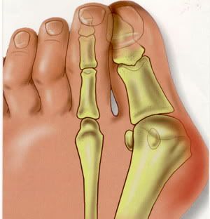 Surgical Treatment Bunion surgery is most often a day case or one night in hospital. Surgery can be done under ankle block (patient awake) or general anaesthetic. It is best to rest with the foot elevated for the first 2 weeks after surgery. The foot is bandaged and a special sandal supplied by the hospital is worn for 6 weeks. Sensible shoes are to be worn for a further 6 weeks after the bandages are removed. It will take between 3-6 months for the swelling to go down. It will take 12 months before everything completely settles. It is also important to remember that not all bunion operations are entirely successful. Overview
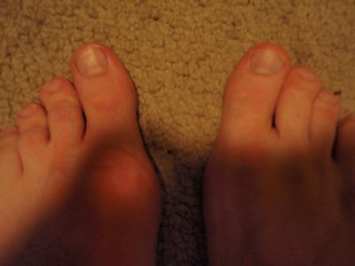 A bunion, also known as hallux valgus, is a painful deformity that develops at the base of the big toe. Bunions are caused when the big toe pushes and bends inward towards the other toes. This displaces the bones of the joint, causing it to protrude in a way that looks like a large growth. Bunions develop due to a variety of factors. Some people inherit feet that are more susceptible due to their shape and structure, having flat feet for instance. But bunions can be made worse by the wrong shoe, or by carrying extra weight or prolonged periods of standing or walking. Causes Bunions are most often caused by an inherited faulty mechanical structure of the foot. It is not the bunion itself that is inherited, but certain foot types that make a person prone to developing a bunion. Although wearing shoes that crowd the toes won?t actually cause bunions, it sometimes makes the deformity get progressively worse. Symptoms may therefore appear sooner. SymptomsA bony bump along the edge of the foot, at the base of the big toe (adjacent to the ball of the foot) Redness and some swelling at or near the big toe joint. Deep dull pain in the big toe joint. Dull achy pain in the big toe joint after walking or a sharp pain while walking. The big toe is overlapping the second toe, resulting in redness, calluses, or other irritations such as corns. Diagnosis Diagnosis begins with a careful history and physical examination by your doctor. This will usually include a discussion about shoe wear and the importance of shoes in the development and treatment of the condition. X-rays will probably be suggested. This allows your doctor to measure several important angles made by the bones of the feet to help determine the appropriate treatment. Non Surgical Treatment Podiatrists will treat bunions conservatively, using paddings and orthotics, which are devices that are made to protect the joint or deviate pressure away from it. Sometimes bunions will develop overlying callus or corns. These can be removed by a podiatrist, but if the area is irritated again by wearing ill-fitting footwear, the corn will grow back. Most people with this condition have flat feet, so arch supports are often recommended.  Surgical Treatment Bunion surgery is most often performed as an out-patient, this means you go home that same day. It will likely be done at a hospital or out-patient (ambulatory) surgery center. The anesthetic choices with bunion surgery are local with sedation, spinal or general anesthesia. You wouldn?t expect that a small bunion would be treated exactly the same as a large one. Over the years, surgeons have developed dozens of methods to surgically correct bunions. But don?t worry because only a handful of methods are used today. With most bunion surgeries today, the procedure involves a combination of soft-tissue rebalancing of ligaments and tendons as well as bone work to re-align the foot structure. You may have heard people say they had their ?bunion shaved.? In most cases, the surgery often involves much more than simply shaving the bunion. The shaving part of the procedure is called an ?exostectomy? and often performed in conjunction with other methods. Overview
 The common bunion is a localized area of enlargement of the inner portion of the joint at the base of the big toe. The enlargement actually represents a misalignment of the big toe joint (metatarsal phalangeal joint) and, in some cases, additional bone formation. The misalignment causes the big toe to point outward (medically termed hallux valgus deformity) toward the smaller toes. This deformity is progressive and will increase with time. The enlarged joint at the base of the big toe (the first metatarsophalangeal joint, or MTP joint) can become inflamed with redness, tenderness, and pain. A small fluid-filled sac (bursa) adjacent to the joint can also become inflamed (bursitis), leading to additional swelling, redness, and pain. A more deep joint pain may occur as localized arthritis develops in later stages of the deformity. A less common bunion is located at the joint at the base of the smallest (fifth) toe. This bunion is sometimes referred to as a tailor's bunion or bunionette. Causes Long periods of pressure from a tight-fitting shoe can cause the inflammation and the pain. This often happens when the big toe is forced into a position where it presses inward and overlaps the second toe. The base of the big toe then is pushed beyond normal alignment of the foot, resulting in the prominence typical of a bunion. SymptomsThe main sign of a bunion is the big toe pointing towards the other toes on the same foot, which may force the foot bone attached to it (the first metatarsal) to stick outwards. Other symptoms may include a swollen, bony bump on the outside edge of your foot, pain and swelling over your big toe joint that's made worse by pressure from wearing shoes, hard, callused and red skin caused by your big toe and second toe overlapping, sore skin over the top of the bunion, changes to the shape of your foot, making it difficult to find shoes that fit. These symptoms can sometimes get worse if the bunion is left untreated, so it's best to see a GP. They'll ask you about your symptoms and examine your foot. In some cases, an X-ray may be recommended to assess the severity of your bunion. Anyone can develop a bunion, but they're more common in women than men. This may be because of the style of footwear that women wear. Diagnosis Your doctor can identify a bunion by examining your foot. Watching your big toe as you move it up and down will help your doctor determine if your range of motion is limited. Your doctor will also look for redness or swelling. After the physical exam, an X-ray of your foot can help your doctor identify the cause of the bunion and rate its severity. Non Surgical Treatment Treatment options are based on the severity of the deformity and symptoms. Nonsurgical treatments usually are enough to relieve the pain and pressure on the big toe. Your doctor may tell you to start wearing roomy, comfortable shoes and use toe padding or a special corrective device that slips into your shoes to push the big toe back into its proper position. To help relieve pain, you can take over-the-counter medications such as acetaminophen (Tylenol) or ibuprofen (Advil, Motrin and others). Whirlpool baths also may help to ease discomfort.  Surgical Treatment If your bunion has progressed to the point where you have difficulty walking, or experience pain despite accommodating shoes, you may need surgery. Bunion surgery realigns bone, ligaments, tendons and nerves so your big toe can be brought back to its correct position. Orthopedic surgeons have several techniques to ease your pain. Many bunion surgeries are done on a same-day basis, requiring no hospital stay, using an ankle-block anesthesia. A long recovery is common and may include persistent swelling and stiffness.
In the last post I suggested that you can DIY certain acupuncture points and treat yourself. Today let's talk about the Five Tigers and how to use them.
There you have it - wearing stilettos or high-heeled shoes for long periods of time can result in foot and ankle problems that include , heel pain, and painful trapped nerves. Cramming toes into narrow pointed-toe shoes can cause corns, calluses and toe deformities. 
One aspect of that junk is uric acid. If your body does not flush this type of acid effectively you will undoubtedly get this type of arthritis. This painful disease is caused when uric acid forms into crystals between joints. A common joint that is typically affected is your Halux Valgus. Bromelain can be purchased in capsule form and taken internally or you can simply eat a lot of pineapple. Pineapple is a tasty way to treat bunions and bruises and also has other natural healthy affects on the body. They asked me if I wanted to talk to a grief counselor. It's a toe people, if it were a foot or leg, then yes, I would definitely need to talk to someone as well as having the med cart by my bedside. At that stage, I still didn't have any pain or discomfort and they started getting worried that I had lost sensation. A few jabs, pokes and pinches later and they were satisfied that I still had feeling in my leg and foot but an unbelievably high pain threshold. Any women can tell you that if you want to experience Foot Pain, walk around for a day in high heels. Heels, combined with fashionable narrow-toed shoes are a leading cause of Foot Pain, as well. Try to avoid these torture machines at all costs, or keep them for special occasion when you know you won't be standing for long periods. Also, replacing your shoes on a regular basis is important, too since even the most expensive shoes break down over time. Attacks of acute gout can be triggered by a number of factors. The aggravating factors include stress, excessive alcohol in the bloodstream, or if you have an underlying medical condition. The painful attacks could last for three to 10 days. Subsequent attacks could be longer and may last for weeks. Remember that the next acute gout attacks will become longer coupled with more intense pains. Never take time to EUR~break in' a pair of new shoes. The time you spend' breaking in' in new shoes could be detrimental to the health of your feet. If a shoe does not feel good the moment you slip it on and walk, you should consider another style. if one shoe hurts your foot, it can cause you to limp. Half a day at work of limping can cause serious lower back pain. Going straight home from work to soak your aching feet each day is never any fun as well. Shoes that rub blisters on your feet can cause intense pain. The bunions you have on your feet can become worse by wearing ill-fitting shoes. Avoid these and many more circumstances surrounding your feet by taking the time to learn more before you shop for your ">discount shoes.
You've just about had it with trying to find a great boot. You want to know the truth about Hi-Tec Altitude IV boots. What you hear about it makes you wonder if this really could be the boot you've been searching for. You've tried all kinds-those purchased at a discount store to those whose cost suggests they're made of gold. You really didn't expect the discount store product to be all that great, but you DID expect the gold-plated one to at least deliver on comfort, being waterproof, keeping your feet warm, and affordable. Alas, such was not the case. Oh, it DID deliver on everything but affordability. So, let's examine the truth about this product.
Some of the more common issues that can be caused by poorly fitting shoes are calluses or even . Calluses, are not usually painful can be unsightly as the skin hardens. Bunions on the other hand can be extremely painful and may need treatment. All of these conditions can be alleviated or even avoided By wearing correct sized shoes. Many women will make the mistake of thinking that by wearing larger sized shoes they will be wide enough for a good, comfortable fit. This is not true, selecting wide womens shoes with the correct width fitting will ease any current problems and help avoid issues in the future.  While most toe deformities are a result of poorly fitting shoes, a claw toe is caused by muscle imbalance, and the imbalance may be due to conditions like rheumatoid arthritis, polio, neuromuscular disorders, a stroke or other conditions. A claw toe is a toe that is contracted at the middle and end joints and can occur in any toe except the Halux Valgus. It can lead to severe pain and pressure. The toe curls downward due to tendons and ligaments that have tightened. The top part of the toe rubs against the shoe causing discomfort at the top and the end of the toe. The toe is also pressed against the bottom of the shoe causing pain. Men who purchased this boot affirm that they are waterproof. Proof of this is dry feet after being submerged in a swamp and two hours of shoveling snow. Hi-Tec's web site, however, does say that the leather does need to be treated regularly to maintain waterproofing. The site gives excellent instructions on how to care for the boot. This terrible situation is made doubly worse when the pointy shoes have heels that are over two inches high. Below two inches is tolerable, but over that is bad news. Then this force called gravity just pushes the foot hard in a real mean way and the foot can't fight back. It's trapped and can't get free. There's no way to get away from all that pressure. Generally, if you have Foot Pain there is something that is not correct. You may have other signs and symptoms. You may have hip, back or knee pain and not connect it to your feet. There's a visual we all need ... thieves, cigarettes, bongs and scales. The question that instantly popped into my mind was: what kind of scales? Were they the kind of scale designed to weigh the substance used in bongs or the kind of scale of the bathroom variety? Although they first became popular for their performance athletic shoes, New Balance now offers a wide range of comfortable styles for the whole family. Let your kids try out a pair when they head back to school.
There are many types of chronic pain and each one differs in degree and severity. Depending on the nature and extent of the pain, most persons who suffer from chronic pain try to manage and treat it by themselves at first. Usually, pain management specialists are consulted only when the pain becomes unbearable.
The flip side to this story is that patients with halux valgus or your tradition bunion can often delay treatment until they start to have pain. in the early stages are a cosmetic concern, but the joint is usually not damaged until the later stages. It is important to address halux valgus when it starts to hurt so the joint is not permanently injured, but a bump, in the absence, of pain can wait. 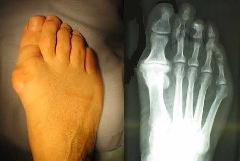 Gout appears because the blood has to much uric acid in it and this acid forms small crystals in the tissues found in the joints. These crystals cause inflammation and pain. The joint that gout mostly affects is the one from the left or right Halux Valgus. Let's say you've chosen a niche. You have chosen to start a rare comic book site. And you have the merchandise. So far it's a vessel that holds niche merchandise. It's not a niche site until you fashion it as the ideal sales and marketing tool for this target market. They asked me if I wanted to talk to a grief counselor. It's a toe people, if it were a foot or leg, then yes, I would definitely need to talk to someone as well as having the med cart by my bedside. At that stage, I still didn't have any pain or discomfort and they started getting worried that I had lost sensation. A few jabs, pokes and pinches later and they were satisfied that I still had feeling in my leg and foot but an unbelievably high pain threshold. Of course since you are young you won't see it at the moment but eventually everything will come at once and the pain will be unbearable under the ball of your foot. This is best known as ball of Foot Pain and this is a very serious problem to deal with and cure. It is very dangerous when you start to feel pain and you think is just temporary and continue to work long hours on your feet. It is sad to say but many people ignore the problem instead of doing something about it, which usually leads to even more problems in the long run. Obtaining a great pair of walking socks are a good method to deter blister formation. The constant rubbing of the foot with fabrics have the possible to produce blisters. Consequently, it's important to keep moisture from soaking the foot throughout activities. If the socks doesn't fit correct, they will bunch up which will improve shearing. Don't worry about whether or not you can afford or whether you have the time to go to physical therapy. You can start treating your foot pain and cure your plantar fasciitis right away, at home. |
AuthorWrite something about yourself. No need to be fancy, just an overview. Archives
July 2017
Categories |
 RSS Feed
